Art meets (health) science in new collaborative anatomy class

Students from the School of the Arts and the Faculty of Health Sciences' Interprofessional Dissection Program gain a new perspective and fresh appreciation for the human form.
BY Sara Laux
April 4, 2023
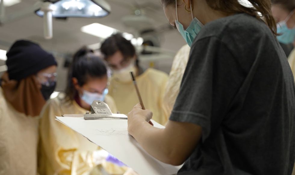
It’s a Tuesday evening. Three McMaster art students are in the anatomy lab in the basement of the Health Sciences Centre, sketching beside some Health Sciences students, who are dissecting a cadaver.
Art students at McMaster have long followed in the footsteps of legendary artists like Leonardo da Vinci and Mansur Ibn Ilyas, drawing preserved specimens of bones, organs, and structures, as well as bodies that have been generously donated for the purposes of education and research.
Now, a more formal arrangement has students from the School of the Arts learning their craft alongside Faculty of Health Sciences students in the interprofessional education (IPE) dissection course.
The collaboration between art and science has seen the students from both faculties not only learning from the specimen in front of them, but also from each other.
Different perspectives and ways of knowing
The IPE dissection course, which is a collaboration between the Faculty of Health Sciences’ Education Program in Anatomy and the Program for Interprofessional Education, Practice and Research (PIPER), is open to students in seven health sciences programs, including medicine, nursing, midwifery and rehabilitation sciences.
Admission to the course is by lottery. And while the 30 Health Sciences students selected do learn anatomy through dissection, there’s another reason that people from vastly different programs all take the same class.
“One of the ultimate goals is getting the students to work with each other and trust each other – because the earlier in their career they can learn that sense of collaboration, the more it will benefit patients further down the line,” explains Yasmeen Mezil, an assistant professor in the department of Pathology and Molecular Medicine, the instructor for the class, and an artist herself.
“We’ve taken the collaboration that already exists among the health sciences students and pulled the art students into it as well,” she says.
Mezil connected with School of the Arts associate professor Briana Palmer to work on incorporating art students more formally into the IPE dissection course.
“I’ve seen a lot of hearts, but I’ve never seen one look like THAT. I’ve never seen such a bold rendering. It captures, not just what the heart looks like, but what it means.” — Bruce Wainman | Director, Education Program in Anatomy
That collaboration extends beyond simply learning anatomy, says Palmer, who coordinates her students’ participation in the classes: three per class, with a signup list each week that regularly fills up.
“This is a way to bring together different ways of looking at things and different ways of knowing,” she says. “This has historically been a very technical space to learn anatomy – but when the health sciences students and the art students share this collective lens, they start to consider the same questions: how do we look at the body? How do we look at life, and how do we express that? What are our roles, and can they move back and forth?”
A once-in-a-lifetime opportunity
For the first 90 minutes or so of every class, the health sciences students present to each other and work on case studies together, while the art students use the vast library of preserved specimens to draw everything from vertebrae to nerves.
After that, while the health sciences students dissect a cadaver, the art students move around them, constantly looking for different perspectives — each discipline learning from the donated body in a slightly different way.
As the final class begins, second-year art student Alysha Aran stands with Bruce Wainman, the director of Mac’s Education Program in Anatomy.
Wainman holds a pelvis in his hands.
“What do you think — male or female?” he grins.
Aran looks thoughtful, and Wainman gives her a hint.
“Remember that a baby’s head is about the size of both your hands clasped together, and that needs to fit through the opening in the pelvis.”
Aran clasps her hands and tries to fit them through the opening between the hip bones. They won’t fit.
“Male,” she says.
Later, as Aran works on sketching the pelvis in big black strokes, the whiff of permanent marker mixes with the lab’s permanent smell of disinfectant.
Her classmate, Lauren Kish, is working on her own drawings, and shares what she’s gained from the class.
“Any new chance to draw, any new setting, any new experience, is important for an artist,” Kish says as she draws. “This is really a once-in-a-lifetime opportunity.”
For the health sciences students, watching the art students’ drawings take shape as a dissection is underway means they get a different perspective on the human body — one that can be clearer than what they see in real life.
“The art students show the anatomy in such a beautiful, but easy-to-understand, functional way,” says Junaid Habibi, a first-year medical student.
“Anatomy can be complicated, and it’s sometimes hard to understand what the different relations are between structures — but when you look at a drawing, then look back at the body, you feel like you get it, and everything makes sense, because they manage to capture it so clearly.”
Savanna Malli, a first-year physiotherapy student who is also an artist, points out that having an art student at her side while she’s dissecting is a good opportunity to share what she knows and solidify her knowledge.
And, Malli says, the learning goes both ways.
“A lot of the art students were really curious about different organs and structures, so I’d be able to tell them, ‘This is a lung, this is what it does, these are the things we’re looking at, and these are the diseases that we might see,” she explains.
“And they would teach us things too — this is how they see the lung. Every body part has its own story, and I think art helps express that.”
A complete change in perspective
The culmination of the class happens a week after the students’ time in the lab has wrapped up. This time, it’s the health sciences students visiting a space that’s familiar to the art students: an open gallery space on the ground floor of Togo Salmon Hall.
The students’ works are displayed on three walls — pages and pages of drawings in pencil, charcoal and marker. Nothing’s framed, because it’s not exactly meant to be a formal exhibition.
Instead, it’s an acknowledgement of everything that the students have experienced together over the past few months — as well as an expression of gratitude and remembrance for the donors with whose bodies the students have become so intimately acquainted.
That’s why, on the fourth wall of the gallery space, there are letters written by the health sciences students to the donors — communicating their appreciation as well as participating in the exhibit itself.
“By having this exhibit, we’re able to remember them as a community,” says Mezil. “We’re bringing together disciplines that are not usually in the same space, and talking about how much they were all able to learn from these teachers together.”
During the opening speeches at the event, a common theme, along with gratitude for the donors, is the sense of having experienced a complete change in perspective.
“I never pictured myself holding a scalpel,” says Hassan Hatoum, a first-year physiotherapy student, to a packed room. “This class changes how we see bodies and interact with them. Lungs aren’t just lungs to me anymore — they’re squishy organs filled with tubes that feel like memory foam.
“We only have one body while we’re on this earth, and we can always call that home.”
Even Wainman, who has been director of the Education Program in Anatomy since 2005, says he’s seeing things in a new way, thanks to the class and its unique collaboration.
“I’ve seen a lot of hearts, but I’ve never seen one look like THAT,” he says, gesturing at the drawings on the wall. “I’ve never seen such a bold rendering. It captures, not just what the heart looks like, but what it means.”
And that’s part of the purpose of art, says Palmer — providing a different way of seeing, a different way of understanding, and giving space for the idea that there are many ways to approach the question of life and death.
“Having that kind of connection between the two groups of students means they’re connecting with another way of knowing,” she says. “Both groups looking through the others’ lens is the fantastic thing about this program and this collaboration.”


