
McMaster researchers create method to detect cases of anemia in archaeological remains
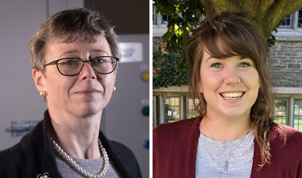
(Left to right) McMaster Professor of Anthropology Megan Brickley and doctoral candidate Brianne Morgan.
BY Wade Hemsworth
March 1, 2024
Diagnosing anemia in living people is typically a matter of a routine blood test.
Retrospectively diagnosing anemia in people who died decades or even centuries ago is much more challenging since there is no blood left to test.
Anthropologists at McMaster University and the University of Montreal, working with a hematologist colleague, have overcome that obstacle by developing a way to detect anemia through patterns in the structures of bones.
Paleopathologists Megan Brickley, who holds the Tier One Canada Research Chair in The Bioarchaeology of Human Disease, and doctoral candidate Brianne Morgan, together with University of Montreal anthropologist Isabelle Ribot and clinician Michelle Zeller, an associate professor of hematology and thromboembolism at McMaster, are the authors of a new study in The Journal of Archaeological Science describing the discovery.
“Anemia is especially prevalent among people of lower socioeconomic status, and it’s likely that was also true in the past,” says Morgan, the paper’s lead author. “Now we have a way we can confirm this with physical evidence.”
The paper describes how the researchers studied the sternum — the plate-like chest bone where the upper ribs meet — and found microscopic gaps between bone layers in living anemia patients subjects which matched patterns in archaeological remains.
Anemia — a deficiency of red blood cells or their components — affects about two billion people worldwide, many of them women and children, causing such symptoms as fatigue, weakness, paleness and shortness of breath.
It’s a significant health concern that was first recognized as a blood condition in the 1800s as the microscope came into common use. Anemia had been recognized well before that time, but only by its symptoms.
Using modern knowledge and technology to understand anemia’s patterns of occurrence through history can help modern physicians and researchers understand more about how and why anemia occurs, especially as it relates to such influences as diet, poverty, sex and age.
“We suspect anemia was really common in the past, but there has been no definitive way to show how common it was,” Brickley says. “Now we can use this completely new approach.”
Red blood cells are made in bone marrow, and it’s possible to diagnose anemia in both living and dead subjects by measuring the gaps between bone layers, which are more pronounced in anemic subjects.
The researchers used micro-CT scanning to study skeletal remains derived from a Quebec cemetery of the 1700s and 1800s and compared them to samples from living patients both with and without anemia to establish the correlation definitively.
The remains are part of a specially designated research collection.
The team examined the microscopic structures of sternum bones, because in life they bear weight less directly than bones from body parts such as the legs, arms and spine. The sternum is less susceptible to fractures and other damage, making structural evidence of anemia easier to isolate.
The researchers studied subjects between 18 and 45, and among the next challenges will be to see whether these gaps may be related to bone fragility, and if the methods can distinguish anemia from osteoporosis in older subjects.

