
Look into your heart: Engineering researchers’ cardiac imaging breakthrough
Zahra Motamed’s lab is developing non-invasive ways for early and precise diagnosis of patients with a chronic cardiovascular disease in which the aortic valve opening narrows, restricting blood flow.
May 1, 2024
McMaster Engineering researchers are developing new, non-invasive ways to diagnose and treat a chronic cardiovascular disease using affordable existing technologies — Doppler and CT scans.
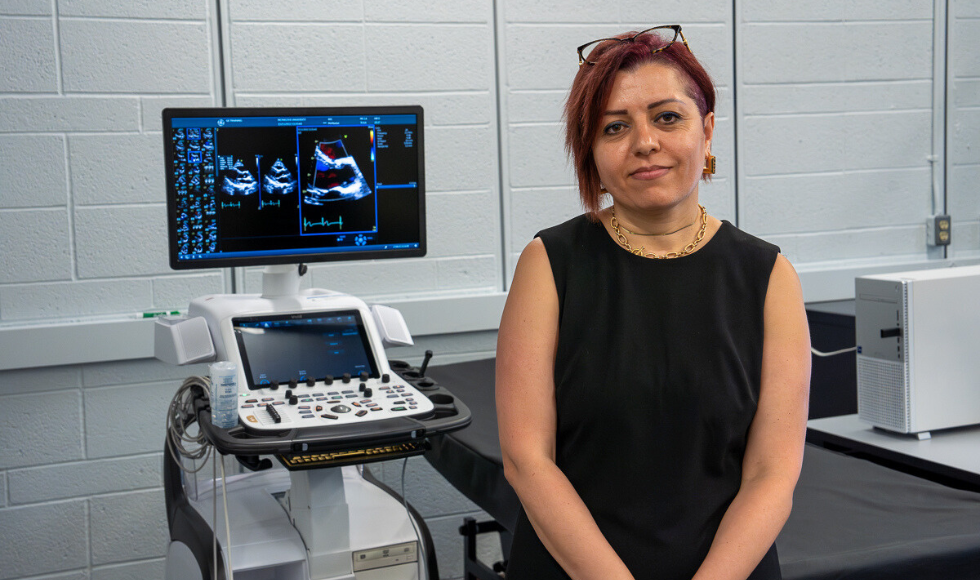
Led by associate professor Zahra Motamed from the department of Mechanical Engineering, the research team has developed two non-invasive ways to diagnose aortic stenosis (AS), a chronic cardiovascular disease, as well as a method of intervention with transcatheter aortic valve replacement (TAVR).
AS is an acute and chronic cardiovascular disease in which the aortic valve opening narrows, restricting blood flow from the heart to the aorta.
Patients with AS are often asymptomatic for a long time before suddenly becoming symptomatic.
Once that happens, patients have a 25-per-cent increase in mortality rate per year, with 50 per cent of symptomatic patients dying within two years if the condition is left untreated, underscoring the need for precise and early detection and intervention.
The Motamed Lab uses Doppler-based tools for diagnosis, qualifying blood flow and valve dynamics, as well as CT-based diagnostic and procedural planning tools for patients with AS who are candidates for TAVR.
“Presently, clinical decisions are based on the emergence of symptoms, which is qualitative and subjective instead of being quantitative,” says Motamed.
Her team has developed two non-invasive diagnostic frameworks: The first uses fully Doppler-based diagnostic tools to quantify blood flow and valve dynamics.
Doppler is the most versatile tool for diagnosis as it is low-cost and risk-free, Motamed notes. “This is incredibly important as patient with cardiovascular diseases need to have regular follow up (on weekly basis or monthly basis etc.).”
The Motamed lab uses Doppler echocardiography to take a series of 2-D images and build an accurate 3-D model of the dynamic behaviour of the heart, says Nikrouz Bahadormanesh, a PhD candidate.
“With this detailed, non-invasive imaging we’re able see how the function of a heart valve is being impaired.”
The second framework they developed uses CT-based diagnostic and procedural planning tools for patients with AS and candidates for transcatheter aortic valve replacement.
“The tools can detect and quantify various properties of aortic valve calcification in addition to reconstructing the 3D model of the patient specific aortic valve,” Motamed says. “None of that is available currently via invasive or non-invasive imaging techniques.”
The CT imaging offers very high field of view — that is, a high level of detail of very small structures — of the entire heart, says PhD candidate Mohamed Abdelkhalek.
“Compared to other imaging methods, CT provides a more accurate geometric picture of the heart that helps to diagnose a disease and manage treatment.”
The CT tools can also detect and quantify aortic valve calcification and reconstruct a 3D model of each patient’s aortic valve, Abdelkhalek notes.
“None of that is available currently with invasive or non-invasive imaging techniques.”
The work has the potential to transform clinical practice and the design of personalized care plans for cardiovascular patients who require regular follow-up care.
“This personalized approach minimizes the potential for complications, reduces health-care costs, and enhances patient care and satisfaction,” says adjunct professor Seyedvahid Khodaei, a postdoctoral fellow in the Motamed Lab.
Motamed notes that while there have been some amazing advancements in medical imaging technology made over the last 10 years, “no one method can give the level of information that we’re able to with what we’re developing here.”
About the researchers
Motamed and her team have worked with clinical collaborators in McMaster’s Faculty of Health Sciences, the University of British Columbia, Hamilton Health Sciences, St Joseph’s Healthcare and the Hospital Universitario Marqués de Valdecilla in Spain.
Her research has been published in the Journal of the American Heart Association, Journal of Medical Image Analysis, JACC: Cardiovascular Imaging, Nature Scientific Reports, Structural Heart, European Heart Journal and REC: Interventional Cardiology (Editorial)

